
How are spinal cord injuries diagnosed?
Authors: SCIRE Community Team | Reviewer: Arlene Aspinall and Tova Plashkes | Published: 17 February 2017 | Updated: ~
Spinal cord injuries are diagnosed using a combination of different assessments and medical tests. Different tests will be chosen by the health team in different circumstances.
Interviews
Interviews with the health team establish the person’s personal and medical history, important details about the injury and what their health needs are.
Neurological assessment
Neurological assessment helps to diagnose problems with the function of the brain, spinal cord, and nerves. This includes physically testing reflexes, sensation, strength, and movement. The most common assessment used for spinal cord injury is the International Standards for Neurological Classification of Spinal Cord Injury (ISNCSCI) exam.
International Standards for Neurological Classification of Spinal Cord Injury Exam (ISNCSCI Exam)
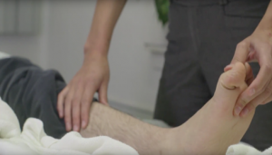
Testing muscle strength as part of the ISCNSCI Exam.5
The ISNCSCI Exam (often called the American Spinal Injury Association (ASIA) Exam) is a physical exam that is used to classify spinal cord injuries. It determines the neurological level of injury and the completeness of the injury.
This exam involves a health provider carefully testing muscle strength, pinprick sensation, and light touch sensation at specific areas on the right and left sides of the body.
Diagnostic imaging and other tests
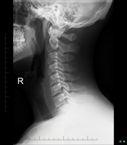
X-ray of the cervical spine (neck).6
X-rays use radiation to visualize the bones of the spine. X-rays can identify broken bones, unstable parts of the spine, and help monitor healing bones. They are also sometimes used to look at possible changes in the lungs or other soft tissues.
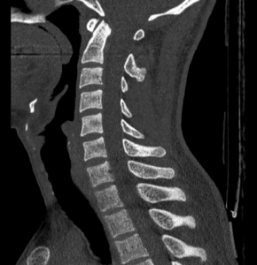
Computed tomography (CT) scan of the cervical spine (neck).7
Computed tomography (CT) uses a series of x-rays taken from different angles that are compiled together to create cross-sectional images that appear as slices through the body. CT can be used to see the bones and tissues in greater detail than x-rays.
Magnetic resonance imaging (MRI) uses a magnetic field and radio waves to create cross-sectional images through the body. MRI is usually used to view the spinal cord and other soft tissues like muscles, nerves, and blood vessels in greater detail.
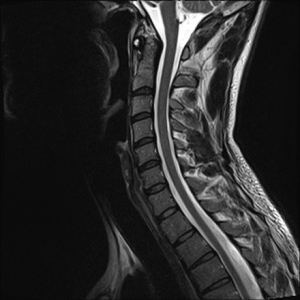
Magnetic resonance imaging (MRI) scan of the cervical spine (neck).8
Electrophysiological testing measures electrical activity in the nerves, spinal cord, and brain. Nerve conduction studies, somatosensory evoked potentials, and motor evoked potentials are types of electrophysiological testing. In these tests, one part of the nervous system, like the brain or the nerves, is stimulated using magnetic or electrical signals and the response elsewhere in the body is then measured. This is used to further understand conduction in the nervous system. It is sometimes used during surgery as well as later in the hospital stay or as an outpatient.
Blood tests study the makeup of the blood to help guide treatment of many different conditions related to SCI.