
How is osteoporosis after SCI diagnosed and measured?
Author: SCIRE Community Team | Reviewer: B. Catharine Craven | Published: 11 October 2017 | Updated: 14 March 2019
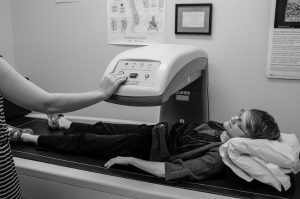
A DXA test is a non-invasive way to measure bone mineral density.8
Measuring and monitoring bone mineral density after SCI is an important part of managing bone health. These measurements allow health providers to diagnose osteoporosis, estimate your fracture risk and monitor changes in your bone density over time. There are several methods that are commonly used to measure different aspects of bone health.
A bone density test or DXA (Dual-Energy X-ray Absorptiometry) Test is an imaging modality that uses non-invasive x-ray technology to scan and measure bone mineral density. DXA is the most widely used tool for measuring bone mineral density and diagnosing osteoporosis.
Peripheral Quantitative Computed Tomography is another non-invasive imaging method that can be used to diagnose osteoporosis of the shin region. This type of scan measures cortical thickness and trabecular volumetric bone mineral density. However, it is most often used in research.
Diagnosing osteoporosis
Health care providers will review your medical history, measure your blood chemistry and bone density, and ask you about your risk factors for fracture before diagnosing osteoporosis or estimating your future risk of fracture. After a fracture, you should undergo screening for osteoporosis.